

Blood is a vital and complex fluid that plays a crucial role in maintaining the health and functionality of the human body. Comprising a combination of red and white blood cells, platelets, and plasma, blood serves various essential functions that are indispensable for life.
The Cardiovascular System
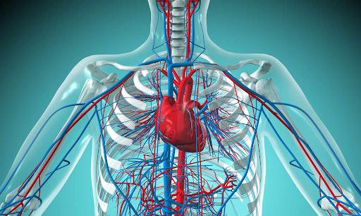
The cardiovascular system is also known as the circulatory system. It transports blood to the lungs in order to send oxygen and blood to the digestive system for nutrients. These essential elements are then delivered throughout the body. In addition, this system collects waste from the body and sends them to the organ systems that remove waste. For example, blood collects urea (a waste product produced by protein breakdown) and circulates it through the kidneys. Here, the urea separates from the blood and leaves the body with urine. In the cardiovascular system, there is the heart and blood vessels. Blood carries oxygen and nutrients to all cells as it travels through blood vessels.
The Vascular System
The vascular system has multiple tubular structures known as blood vessels. These tubes all come in different sizes, patterns, and designs, but they connect together to form a closed circuit in order to help the heart circulate blood throughout the body. The heart transports blood throughout the entirety of the vascular system, pumping oxygenated and deoxygenated blood. In the heart, there is a wall of muscle called the septum. The septum divides the heart into a left and right half; the right side carries deoxygenated blood while the left side carries oxygenated blood. There is also a second wall, which separates the round upper section of the heart from the cone-shaped lower section. In total, there are four sections, or chambers, of the heart; the upper chambers are called atria and the lower chambers are called ventricles.
Blood Vessels
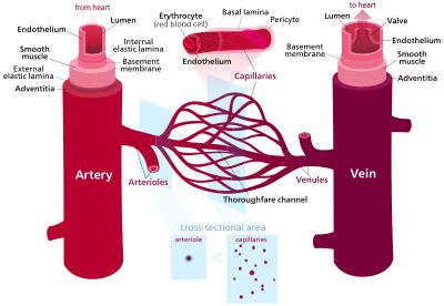
Blood vessels are specifically designed to perform different functions, which is why they all look different. Arteries and veins both consist of the following three tissue layers:
- Tunica intima - inner, smooth layer in direct contact with blood
- Tunica media - middle, thicker layer able to contract and relax
- Tunica adventitia - outer layer. a covering that protects and supports veins
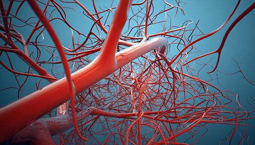
Capillaries are the smallest blood vessels, and they have only one layer of tissue. In fact, they are so small that blood cells have to travel in a single-file line in order to pass through. Arteries, on the other hand, are a bit different because they carry blood away from the heart instead of carrying it to the heart like capillaries. The walls of arteries are flexible but also much thicker than the walls of veins and capillaries because they need to be able to withstand the pressure and intensity of pumping blood from the heart. When the heart “beats”, it pushes blood through the arteries at a high pressure. Think of how high the pressure is when you can hear your heart contracting! This is why you can feel your pulse at specific points on the body, including the radial pulse in the wrist and the carotid pulse in the neck. Since most arteries carry oxygenated blood, arterial blood is bright red.
All arteries except the pulmonary arteries contain oxygenated blood, and all veins except the pulmonary veins carry deoxygenated blood. Pulmonary arteries carry deoxygenated blood from the heart to the lungs, and pulmonary veins carry oxygenated blood from the lungs back to the heart. The aorta is the largest artery, and it carries blood that is rich in oxygen from the heart by branching out into smaller arteries that divide into even smaller arteries until they reach capillaries. As established before, this is a necessary process because capillaries are extremely small. Capillaries act as connection points between arteries and veins (small veins), so blood cells need to be able to pass through them. After carrying oxygen from the blood to the tissues, they merge into veins. Finally, the largest veins – the superior and inferior vena cava – return deoxygenated blood into the right atrium of the heart.
The Heart
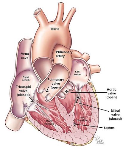
The heart consists of three layers. The inner layer is the endocardium. It is lined with endothelial cells, which are similar to the lining of blood vessels. Cardiac muscle is the muscular middle layer. The left ventricle has the thickest heart muscle because it is responsible for pumping blood to the rest of the body. The outermost layer is the epicardium. This layer consists of two membranes: an internal serous visceral membrane attached to the heart and an external fibrous parietal membrane. These two membranes are separated by pericardial fluid, which is secreted by the visceral (serous) membrane. The two membranes and the fluid inside them are called the pericardium. Blood flows from the atria down into the ventricles because of the holes in the walls that separate the ventricles. These openings are called valves because they open in one direction - like hatches - to let blood through. They then close to prevent blood from flowing back into the atrium. The valve between the right atrium and right ventricle is the tricuspid valve. The valve between the left atrium and left ventricle is the mitral valve (bicuspid). This system allows blood to flow in only one direction within the heart.
Phlebotomy
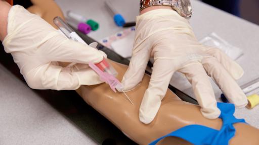
Professionals called phlebotomists make an incision into the skin and blood vessels. The results of laboratory testing are extremely important in providing appropriate, quality healthcare. The primary role of a phlebotomist is to obtain blood specimens for this diagnostic testing. They are tested for everything from levels of glucose, proteins, and drugs to blood cell counts, antibodies, and infectious diseases. Blood is obtained either by venipuncture (puncturing a vein) or dermal (capillary) puncture (puncturing the skin).
An average adult has 8 to 12 pints of blood. A pint is roughly the same as a unit of blood. A unit of blood is what is transfused to a patient when he needs blood. This is the usual amount of blood given during a transfusion. Blood is constantly distributed along more than 70,000 miles of tubes (vessels), collectively called the vasculature. A phlebotomist must understand the circulatory system and its composition and functions. In addition to the cellular and fluid composition of the blood, the phlebotomist must also understand how the closed circuit of blood vessels transports blood. To make a vein point, it is important to know the location of blood vessels, especially the most used vessels, and the composition of the blood.
Medical Laboratory Personnel

There are several professionals in the medical laboratory with whom the phlebotomist may interact, including the following:
- Medical office greets and assists outpatients requiring laboratory services.
- Medical transcription specialists prepare reports dictated by a pathologist.
- Medical laboratory assistants (MLAs) are phlebotomists trained to perform minor tests or otherwise assist laboratory staff.
- Histology technicians (HTs) prepare small sections of surgical specimens for microscopic examination by a pathologist.
- Histologists (HTL) perform the more complex tasks of the histology laboratory and are responsible for the technical aspects of the histology laboratory and for the evaluation of new procedures.
- Cytologists (CT) perform a microscopic examination of human cells to detect cancer and other diseases. In addition, some laboratories employ specialists with master’s or doctorate degrees in certain fields, such as microbiology or molecular pathology.
- Pathologists are doctors who specialize in the study of disease, which includes anatomical and clinical pathology. An anatomical pathologist makes a diagnosis based on surgically removed tissue. A clinical pathologist controls the interpretation of the results of analyzes of blood and body fluids obtained by MLT and MLS. Some pathologists also have specialties in various medical laboratory specialties.
- Pathologists’ Assistants (PAs) examine surgically removed tissue specimens and collect and examine autopsy specimens. They help pathologists identify diseases.
- Medical laboratory technicians (MLTs), formerly called clinical laboratory technicians by some institutions, hold at least an associate degree and can perform low-complexity and some moderately complex laboratory tests except cytology and histology. In some states, MLTs may perform some procedures (require interpretation) under the direct supervision of medical laboratory investigators.
- Medical laboratory scientists (MLS), formerly called medical technologists or clinical laboratory scientists, have at least a bachelor’s degree and can perform complex tests other than cytology and histology. They are responsible for the technical aspects of the medical laboratory and for the evaluation of new procedures. MLSs also have specializations in one or more laboratory departments. Some laboratories employ individuals with degrees in clinical chemistry or medical microbiology for MLS positions in certain parts of the medical laboratory.